Nandrolone for Mood | Feeling much Better..

Buy Lab Tests Online
Also, I haven't read the mouse study, but it occurs to me that in the early days of Low Carb, there was an attempt to show that (I think) olive oil was toxic using FMD as a measure. However I believe Volek subsequently showed that it was a short-term affect which disappeared after several weeks of adaptation. My memory is hazy on the details but the clear takeaway I do remember is that in some cases that the body can adapt to what initially appears to be a problem, so if such an adaptation period was not part of the mouse study, that would call it into question and could explain the apparent lack of issue in humans. There are a number of examples of this that come into play when people transition from a low-fat to a low-carb way of eating.
 
Defy Medical TRT clinic doctor
Also, I haven't read the mouse study, but it occurs to me that in the early days of Low Carb, there was an attempt to show that (I think) olive oil was toxic using FMD as a measure. However I believe Volek subsequently showed that it was a short-term affect which disappeared after several weeks of adaptation. My memory is hazy on the details but the clear takeaway I do remember is that in some cases that the body can adapt to what initially appears to be a problem, so if such an adaptation period was not part of the mouse study, that would call it into question and could explain the apparent lack of issue in humans. There are a number of examples of this that come into play when people transition from a low-fat to a low-carb way of eating.

Rodent Free #cuttheshit for @DS3

@madman
@Nelson Vergel

Interesting study in natal human females that are TGM:


1651167302838.png


1651167336739.png

1651167360395.png


DISCUSSION

We demonstrated for the first time that physiological testosterone administration during HT was associated with impaired endothelial function in young TGM compared to CGF women. Our findings are consistent with earlier data showing endothelium mediated vasodilation is attenuated in lean, insulin-sensitive women with AE-PCOS with endogenous hyperandrogenism11. Importantly, the difference in endothelial function between TGM and CGF was found despite comparable levels of traditional cardiovascular risk factors including BMI, lipid levels and blood pressure, suggesting that other mechanisms such as inflammation or the direct effect of androgens may contribute to changes in endothelial function. There was a small but statistically significant difference in HbA1C in the TGM (5.0 vs 4.7%, P=0.048), however the levels were well within the normal range and unlikely to have clinically relevant impact on vascular function. However, we cannot entirely exclude the possibility that subtle changes in cardiometabolic measurements may have a deleterious effect on endothelial function with long-term treatment. Earlier studies have shown adverse effects of testosterone on lipid levels and blood pressure17, 24, raising concern for increased cardiovascular risk with chronic testosterone exposure. Therefore, attention to cardiometabolic risk factors should be integral to the care of TGM. Finally, because of experimental constraints, our laboratory tests were performed by different laboratories so we were unable to directly compare lipids and hormones between the two groups so these studies should be repeated with blood analysis performed using the same assays for both groups.

Endothelial dysfunction constitutes “the early pivotal event in atherosclerosis”25, because it precedes clinically detectable atherosclerotic plaques in the coronary arteries. Healthy endothelial function may be described as a balance of endothelial vasoconstrictor/vasodilator factors. A primary feature of normally functioning endothelium is the production of nitric oxide in response a number of different agonists (e.g. shear stress in FMD). Testosterone exposure is also associated with impaired agonist-triggered endothelial NO release in women3, 4, and is therefore a major driver of endothelial dysfunction. Activation of the AR in women may result in impaired, agonist-triggered endothelial NO release and/or NO responsiveness. A recent study in our laboratory demonstrated impaired endothelin-1 (agonist) triggered vasodilation in lean, insulin sensitive women with AE-PCOS, indicating an independent effect of androgen on vascular pathology in these women with high testosterone exposure but no insulin resistance or obesity11. This same study indicated the endothelial dysfunction in AE-PCOS is mediated through an androgen effect on the NO pathway11. Earlier data in PCOS had demonstrated that inflammation and inflammatory factors including cytokines, oxidative stress and NF-κB activation26 also contribute to the impaired NO release or responsiveness5, 6. These data are consistent with our current findings that the impaired endothelial function in TGM is independent of obesity or blood lipids and likely associated with inflammatory factors associated with the testosterone exposure.


Testosterone is an acute vasodilator and in men may protect against endothelial dysfunction27. Androgen receptors are expressed in cells throughout the cardiovascular system, including endothelial cells28 and vascular smooth muscle cells (VSMCs)29, although the impact of testosterone administration on the cardiovascular system in men is varied2, 3033. In contrast, androgens may induce detrimental outcomes on the cardiovascular system in women24. In general, the engagement of androgen and the androgen receptor results in impaired, agonist-triggered endothelial NO release in women, a likely cause of the sex differences in testosterone effects on endothelial function. A recent meta-analysis of androgen (DHT) treatment showed dyslipidemia at 3, 6 and 24 months of testosterone treatment in transmen17. While HT with testosterone also induced blood pressure increases, insulin resistance, and dyslipidemia17, cardiovascular morbidity or mortality was not yet apparent in these young transgender men24. No studies have yet followed transgender men into the aging process as cardiovascular disease risks accelerate, therefore the long-term impact on cardiovascular disease remains unknown.

Earlier studies have shown Hb and Hct during testosterone treatment (in men) can increase iron and red blood cell formation34, and that Hb is inversely related to forearm endothelial-dependent vasodilation35. However, in this study, while Hct was greater in the TGM versus the CGF, there was no relationship within either group to FMD or when we considered the groups as a whole. Further studies examining this specific relationship are required as our blood samples and FMD measures in TGM were not taken on the same day, and Hb and Hct are sensitive to hydration and posture, among other variables.


In women, [TTotal]S is associated with greater risk of diabetes and related cardiovascular comorbidities37. In the present study, we noted small but significantly greater HbA1C in the TGM (5.0 vs 4.7, P=0.048) in this young, healthy cohort. In this group we would expect the incidence of impaired glucose metabolism to be very low. The normal range for HbA1C is <5.7%, prediabetes is defined as an HbA1C 5.7–6.4%, and diabetes is defined as an HbA1C ≥ 6.5%. All subjects in both groups were in the normal range (4.0–5.6%), suggesting while this was statically different, there is little physiological or clinical consequence.

Conclusions

Ours is the first study to examine the androgen effects on endothelial function in TGM. We demonstrated that the hyperandrogenic milieu in TGM is a primary factor associated with endothelial dysfunction, independent of lipids, blood pressure and BMI. Our present study supports earlier studies from our laboratory demonstrating that poor NO responsiveness is a key causative link in natal females exposed to chronic endogenous and/or high levels of exogenous androgens leading to endothelial dysfunction and ultimately cardiovascular disease. Future studies should address how changes in other hormones such as estrogen, or other substances, such as inflammatory cytokines, may impact changes in vascular function during HT. As described earlier, endothelial dysfunction in these TGM occurred independent of differences in lipids, BMI or blood pressure indicating a separate etiology. Understanding the mechanisms by which exogenous androgens mediate endothelial dysfunction in TGM may allow for early interventions to mitigate the potential long-term cardiovascular risk.


As pointed out in the Hct thread, there's obviously a limit for each individual how much TT they can handle before FMD/endothelial function becomes impaired rather than enhanced (important in the context of Hct elevation, blood viscosity, shear stress). Hence, u-shaped curve for TT on morbiditiy just like most other hormones, co-factors, etc.
 
Last edited by a moderator:

1651169069664.png


1651169087002.png


No rodents here...


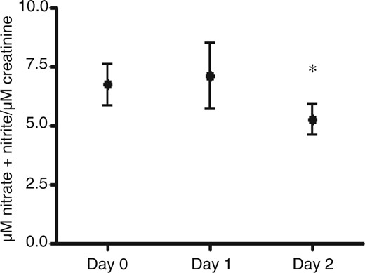
Here we show that supra-physiological concentrations of testosterone inhibit the urinary excretion of NO in healthy volunteers 48 hours after the administration of testosterone. Since urinary NO is a biomarker for endothelial function and mainly originates from the endothelium, our results indicate that even a single dose of testosterone may induce a decrease in NO formation in endothelial cells. One determinant of NO bioavailability is the expression and activity of eNOS, the main enzyme involved in the metabolism of NO in endothelial cells. Here we show that testosterone down-regulates the gene expression of eNOS after 48 hours. In a previous study it was shown that low doses of testosterone induce the protein expression and activity of eNOS in HUVECs and in rat aorta. These effects were lost when higher doses of testosterone were used, and in agreement with our findings, an inhibition in eNOS expression and activity could be discerned.16

Since addition of the antioxidant SeMe partly abolished the inhibitory effect of testosterone on eNOS gene expression, we speculate that oxidative stress may at least to some extent explain the transcriptional inhibition of eNOS and subsequently the NO production. Therefore we further studied whether supra-physiological doses of testosterone induce oxidative stress in vivo. Indeed, this turned out to be right as the results revealed an inhibition of the antioxidative capacity in vivo probably by generation of reactive oxygen species (ROSs) and/or inhibition of the antioxidative activity. This would be in line with our in vitro results where several important defence enzyme genes, that is, CAT, SOD1 and GPX4, were down-regulated by a supra-physiological dose of testosterone. The decreased transcriptional activity was observed already after 8 h exposure, indicating that oxidative stress may be acutely induced by testosterone. However, after 48 h the mRNA expression levels of these enzyme genes were back to baseline levels or increased, possibly as a consequence of the induced oxidative pressure in the endothelial cells. Our results indicate that the oxidative stress status was induced by testosterone both in the in vivo situation and in vitro. A previous study in rats has shown that AAS induce oxidative stress. After eight weeks of stanozolol treatment the serum levels of thiobarbituric acid-reactive substances (TBARSs) increased.17

It is well known that long term AAS abuse has detrimental effects on the cardiovascular system, to which the increase of LDL and decrease of HDL may contribute. However, many adverse side-effect observations are based on anecdotal evidences and the available scientific data are scarce. It is therefore important to scientifically identify and characterize the side-effects of AASs.18 Despite the liability of AAS abusers to develop cardiovascular disorders the AAS-related effects on the endothelium have received little attention. D'Ascenzo et al. demonstrated that exposure of HUVECs to AAS alters endothelial cell growth with a strong anti-proliferative effect, induces apoptosis and modifies intracellular levels of calcium.9 Two studies have observed that the flow mediated dilatation of the brachial artery determined by ultrasound (an in vivo index of endothelial function) was reduced in body builders using AASs.8,19


The baseline serum testosterone concentration in the study population was 5 ng/ml.20 This is the same as has been seen in other study groups similar in respect of age, gender and ethnicity.21 Two days after testosterone administration the serum testosterone level has increased by 200%. The concentration used in our in vitro experiments (1 µM) is in the range of the levels achieved after administration of testosterone to the volunteers.20 In this study we have expressly studied supra-physiological doses of testosterone in order to mimic AAS-abuse. It is important to stress that testosterone exerts the opposite effect when therapeutic doses of it are used in hypogonadal men. Male hypogonadism has been associated with endothelial dysfunction22 and the decline in testosterone has been associated with metabolic syndrome.23 Testosterone replacement therapy (TRT) has been shown to have beneficial effects on the endothelium.24

It is important to address the limitations of this study since it was conducted in relatively few individuals and after only a single testosterone dose. The results observed here do not necessarily imply that repeated AAS abuse exerts these detrimental effects, or even worsens endothelial function. It is possible that chronic adaptations develop to counterbalance the increased level of oxidative stress or the acute reduction of eNOS mRNA and NO production. The data need to be verified in other studies. However, it is difficult to study the adverse side-effects of AAS abuse in humans because of the ethical restraints limiting the amount of administered testosterone. Retrospective studies from self-reported AAS abusers are sometimes indicative but have a lot of limitations, such as co-administration of other drugs, purity and identity of the AASs, etc.


Rodent Free, #cuttheshit for @DS3

FYI @Cataceous

@Guided_by_Voices Sure would be fun to follow this experiment (~2000 ng/dl TT) out a few years and look at transfer function between micro and macro toxicity for my graph. Makes me rethink how to present the data.

 
Last edited by a moderator:

Reactive oxygen species: players in the cardiovascular effects of testosterone​


Summary

The role of testosterone in mediating or protecting against ROS and antioxidant capacity in the cardiovascular system is far from being clear, with testosterone presenting prooxidant as well as antioxidant effects. There are various possible contributing factors to these discrepant effects of testosterone in the cardiovascular system and in the other tissues and organs: 1) acute vs. chronic differential effects are possibly due to activation of distinct sets of signaling pathways, as reported with other hormones and neurotransmitters; 2) the initial metabolic-energetic-redox status of the cell may exacerbate cardiovascular risk; 3) global (or local) increases in testosterone may produce differential effects based on the specific cell types that are stimulated; 4) the concentrations of testosterone (physiological, supraphysiological) may produce different effects; 5) the steroid ester used (testosterone cypionate, decanoate, undecanoate, enanthate, propionate, heptylate, caproate, phenylpropionate, isocaproate, acetate) changes the compound solubility in water and slows the release of the parent steroid, i.e., changes absorption time. Different esters are also susceptible to the presence of native and selective esterases in the many complex biological/cellular environments and can mask specific functional groups. These processes may confer different responses to different esters; 6) the “sex” of the individual or cell/tissue may determine differential effects of testosterone; and not least, 7) different animal species (mice, rats, humans, rabbits, birds) and the age, duration, and characteristics of the diseases/conditions upon which testosterone effects are determined are not fully understood. The complexity of testosterone effects is evident, and further basic and clinical studies are required for a better understanding of the mechanisms by which testosterone gains its biological activity independent of reproduction, which may be detrimental and/or beneficial to the cardiovascular system.
 
Last edited by a moderator:


Testosterone and dihydrotestosterone modulate the redox homeostasis of endothelium​


Abstract​


The predominance of cardiovascular diseases among men compared to premenopausal women has been attributed to testosterone, which is implicated in vascular remodeling. Molecular mechanisms underlying its role have not been clarified but oxidative stress-induced inflammation may be important. We therefore investigated in vitro the effects of testosterone and dihydrotestosterone, (a nonaromatized androgen), on redox homeostasis in absence (basal conditions) and after corticotropin-releasing hormone-induced pro-oxidant action in macroendothelial cells. More specifically, we explored their role on well-established antioxidant enzymes activity, namely endothelial nitric oxide synthase, superoxide dismutase, catalase, and glutathione. We observed that both androgens significantly increased the intracellular reactive oxygen species levels, endothelial nitric oxide synthase activity, nitric oxide concentration as well as superoxide dismutase activity and decreased catalase activity. These effects of Testosterone and DHT were reversed in the presence of the androgen receptor antagonist, flutamide. Moreover, testosterone and dihydrotestosterone similarly enhanced the stimulatory effect of corticotropin-releasing hormone on intracellular reactive oxygen species levels and superoxide dismutase activity but did not influence the inhibitory effect on endothelial nitric oxide synthase activity, nitric oxide release and catalase activity. Finally, androgens did not have a detectable effect on glutathione levels or the glutathione/glutathione plus glutathione disulfide ratio. Our results reveal that testosterone and DHT rise the intracellular redox threshold of the endothelial cell and increases NO synthesis. These findings suggest that the action of testosterone is affected by the redox status of the endothelium and help to explain its controversial effects on the cardiovascular system.
 
Last edited by a moderator:
Rodent Free #cuttheshit for @DS3

@madman
@Nelson Vergel

Interesting study in natal human females that are TGM:


View attachment 21464


View attachment 21465
View attachment 21466


DISCUSSION

We demonstrated for the first time that physiological testosterone administration during HT was associated with impaired endothelial function in young TGM compared to CGF women. Our findings are consistent with earlier data showing endothelium mediated vasodilation is attenuated in lean, insulin-sensitive women with AE-PCOS with endogenous hyperandrogenism11. Importantly, the difference in endothelial function between TGM and CGF was found despite comparable levels of traditional cardiovascular risk factors including BMI, lipid levels and blood pressure, suggesting that other mechanisms such as inflammation or the direct effect of androgens may contribute to changes in endothelial function. There was a small but statistically significant difference in HbA1C in the TGM (5.0 vs 4.7%, P=0.048), however the levels were well within the normal range and unlikely to have clinically relevant impact on vascular function. However, we cannot entirely exclude the possibility that subtle changes in cardiometabolic measurements may have a deleterious effect on endothelial function with long-term treatment. Earlier studies have shown adverse effects of testosterone on lipid levels and blood pressure17, 24, raising concern for increased cardiovascular risk with chronic testosterone exposure. Therefore, attention to cardiometabolic risk factors should be integral to the care of TGM. Finally, because of experimental constraints, our laboratory tests were performed by different laboratories so we were unable to directly compare lipids and hormones between the two groups so these studies should be repeated with blood analysis performed using the same assays for both groups.

Endothelial dysfunction constitutes “the early pivotal event in atherosclerosis”25, because it precedes clinically detectable atherosclerotic plaques in the coronary arteries. Healthy endothelial function may be described as a balance of endothelial vasoconstrictor/vasodilator factors. A primary feature of normally functioning endothelium is the production of nitric oxide in response a number of different agonists (e.g. shear stress in FMD). Testosterone exposure is also associated with impaired agonist-triggered endothelial NO release in women3, 4, and is therefore a major driver of endothelial dysfunction. Activation of the AR in women may result in impaired, agonist-triggered endothelial NO release and/or NO responsiveness. A recent study in our laboratory demonstrated impaired endothelin-1 (agonist) triggered vasodilation in lean, insulin sensitive women with AE-PCOS, indicating an independent effect of androgen on vascular pathology in these women with high testosterone exposure but no insulin resistance or obesity11. This same study indicated the endothelial dysfunction in AE-PCOS is mediated through an androgen effect on the NO pathway11. Earlier data in PCOS had demonstrated that inflammation and inflammatory factors including cytokines, oxidative stress and NF-κB activation26 also contribute to the impaired NO release or responsiveness5, 6. These data are consistent with our current findings that the impaired endothelial function in TGM is independent of obesity or blood lipids and likely associated with inflammatory factors associated with the testosterone exposure.


Testosterone is an acute vasodilator and in men may protect against endothelial dysfunction27. Androgen receptors are expressed in cells throughout the cardiovascular system, including endothelial cells28 and vascular smooth muscle cells (VSMCs)29, although the impact of testosterone administration on the cardiovascular system in men is varied2, 3033. In contrast, androgens may induce detrimental outcomes on the cardiovascular system in women24. In general, the engagement of androgen and the androgen receptor results in impaired, agonist-triggered endothelial NO release in women, a likely cause of the sex differences in testosterone effects on endothelial function. A recent meta-analysis of androgen (DHT) treatment showed dyslipidemia at 3, 6 and 24 months of testosterone treatment in transmen17. While HT with testosterone also induced blood pressure increases, insulin resistance, and dyslipidemia17, cardiovascular morbidity or mortality was not yet apparent in these young transgender men24. No studies have yet followed transgender men into the aging process as cardiovascular disease risks accelerate, therefore the long-term impact on cardiovascular disease remains unknown.

Earlier studies have shown Hb and Hct during testosterone treatment (in men) can increase iron and red blood cell formation34, and that Hb is inversely related to forearm endothelial-dependent vasodilation35. However, in this study, while Hct was greater in the TGM versus the CGF, there was no relationship within either group to FMD or when we considered the groups as a whole. Further studies examining this specific relationship are required as our blood samples and FMD measures in TGM were not taken on the same day, and Hb and Hct are sensitive to hydration and posture, among other variables.


In women, [TTotal]S is associated with greater risk of diabetes and related cardiovascular comorbidities37. In the present study, we noted small but significantly greater HbA1C in the TGM (5.0 vs 4.7, P=0.048) in this young, healthy cohort. In this group we would expect the incidence of impaired glucose metabolism to be very low. The normal range for HbA1C is <5.7%, prediabetes is defined as an HbA1C 5.7–6.4%, and diabetes is defined as an HbA1C ≥ 6.5%. All subjects in both groups were in the normal range (4.0–5.6%), suggesting while this was statically different, there is little physiological or clinical consequence.

Conclusions

Ours is the first study to examine the androgen effects on endothelial function in TGM. We demonstrated that the hyperandrogenic milieu in TGM is a primary factor associated with endothelial dysfunction, independent of lipids, blood pressure and BMI. Our present study supports earlier studies from our laboratory demonstrating that poor NO responsiveness is a key causative link in natal females exposed to chronic endogenous and/or high levels of exogenous androgens leading to endothelial dysfunction and ultimately cardiovascular disease. Future studies should address how changes in other hormones such as estrogen, or other substances, such as inflammatory cytokines, may impact changes in vascular function during HT. As described earlier, endothelial dysfunction in these TGM occurred independent of differences in lipids, BMI or blood pressure indicating a separate etiology. Understanding the mechanisms by which exogenous androgens mediate endothelial dysfunction in TGM may allow for early interventions to mitigate the potential long-term cardiovascular risk.


As pointed out in the Hct thread, there's obviously a limit for each individual how much TT they can handle before FMD/endothelial function becomes impaired rather than enhanced (important in the context of Hct elevation, blood viscosity, shear stress). Hence, u-shaped curve for TT on morbiditiy just like most other hormones, co-factors, etc.
Not sure what to make of this. It would have been interesting to see a control group of biological men with similar T levels. Also, the Hyperlipid blog had a post some years back about FMD being a far-from-precise measurement. Finally, as I've written about before regarding Nandrolone, there are benefits that may cancel out any negatives. In my case I am able to maintain a significantly higher level of activity on TRT+ that I can without it ( I used to come off for 3 months or so a year so I have a fair amount of experience experimenting.) The higher capacity (due primarily to better recovery capacity) let's me do more of heart-healthy things like sprinting and other conditioning work that would be a recipe for overtraining otherwise, so I hesitate to put too much weight on any one variable.
 
Not sure what to make of this. It would have been interesting to see a control group of biological men with similar T levels. Also, the Hyperlipid blog had a post some years back about FMD being a far-from-precise measurement. Finally, as I've written about before regarding Nandrolone, there are benefits that may cancel out any negatives. In my case I am able to maintain a significantly higher level of activity on TRT+ that I can without it ( I used to come off for 3 months or so a year so I have a fair amount of experience experimenting.) The higher capacity (due primarily to better recovery capacity) let's me do more of heart-healthy things like sprinting and other conditioning work that would be a recipe for overtraining otherwise, so I hesitate to put too much weight on any one variable.
Thanks for your great point. Would have been great to carry out the study (I tagged for you) longer as well.

I fully agree with your point about cumulative net effect. If AAS can get a guy to lose weight and be more active then perhaps he's in a better position than without them. For example, AAS may increase Hct (which increases WBV), but through diet and exercise inflammation may come down resulting in lower plasma viscosity and overall net lower whole blood viscosity (just an example). They definitely helped me with QOL given my issues until I ran into another issue of indeterminate cause.

Clearly there are plenty of folks walking around earth who have years of cumulative use (and some abuse) of AAS. And then we also have anecdotal treasures like @BigTex and others who are the genetic equivalent of 7'6'' tall persons :) . Way to go @BigTex. It was like a guy I was talking to and we were discussing how expensive his hobby was becoming. But he shrugged it off saying it was like oxygen and of course you have to breathe.
 
Last edited by a moderator:
Rodent Free #cuttheshit for @DS3

@madman
@Nelson Vergel

Interesting study in natal human females that are TGM:


View attachment 21464


View attachment 21465
View attachment 21466


DISCUSSION

We demonstrated for the first time that physiological testosterone administration during HT was associated with impaired endothelial function in young TGM compared to CGF women. Our findings are consistent with earlier data showing endothelium mediated vasodilation is attenuated in lean, insulin-sensitive women with AE-PCOS with endogenous hyperandrogenism11. Importantly, the difference in endothelial function between TGM and CGF was found despite comparable levels of traditional cardiovascular risk factors including BMI, lipid levels and blood pressure, suggesting that other mechanisms such as inflammation or the direct effect of androgens may contribute to changes in endothelial function. There was a small but statistically significant difference in HbA1C in the TGM (5.0 vs 4.7%, P=0.048), however the levels were well within the normal range and unlikely to have clinically relevant impact on vascular function. However, we cannot entirely exclude the possibility that subtle changes in cardiometabolic measurements may have a deleterious effect on endothelial function with long-term treatment. Earlier studies have shown adverse effects of testosterone on lipid levels and blood pressure17, 24, raising concern for increased cardiovascular risk with chronic testosterone exposure. Therefore, attention to cardiometabolic risk factors should be integral to the care of TGM. Finally, because of experimental constraints, our laboratory tests were performed by different laboratories so we were unable to directly compare lipids and hormones between the two groups so these studies should be repeated with blood analysis performed using the same assays for both groups.

Endothelial dysfunction constitutes “the early pivotal event in atherosclerosis”25, because it precedes clinically detectable atherosclerotic plaques in the coronary arteries. Healthy endothelial function may be described as a balance of endothelial vasoconstrictor/vasodilator factors. A primary feature of normally functioning endothelium is the production of nitric oxide in response a number of different agonists (e.g. shear stress in FMD). Testosterone exposure is also associated with impaired agonist-triggered endothelial NO release in women3, 4, and is therefore a major driver of endothelial dysfunction. Activation of the AR in women may result in impaired, agonist-triggered endothelial NO release and/or NO responsiveness. A recent study in our laboratory demonstrated impaired endothelin-1 (agonist) triggered vasodilation in lean, insulin sensitive women with AE-PCOS, indicating an independent effect of androgen on vascular pathology in these women with high testosterone exposure but no insulin resistance or obesity11. This same study indicated the endothelial dysfunction in AE-PCOS is mediated through an androgen effect on the NO pathway11. Earlier data in PCOS had demonstrated that inflammation and inflammatory factors including cytokines, oxidative stress and NF-κB activation26 also contribute to the impaired NO release or responsiveness5, 6. These data are consistent with our current findings that the impaired endothelial function in TGM is independent of obesity or blood lipids and likely associated with inflammatory factors associated with the testosterone exposure.


Testosterone is an acute vasodilator and in men may protect against endothelial dysfunction27. Androgen receptors are expressed in cells throughout the cardiovascular system, including endothelial cells28 and vascular smooth muscle cells (VSMCs)29, although the impact of testosterone administration on the cardiovascular system in men is varied2, 3033. In contrast, androgens may induce detrimental outcomes on the cardiovascular system in women24. In general, the engagement of androgen and the androgen receptor results in impaired, agonist-triggered endothelial NO release in women, a likely cause of the sex differences in testosterone effects on endothelial function. A recent meta-analysis of androgen (DHT) treatment showed dyslipidemia at 3, 6 and 24 months of testosterone treatment in transmen17. While HT with testosterone also induced blood pressure increases, insulin resistance, and dyslipidemia17, cardiovascular morbidity or mortality was not yet apparent in these young transgender men24. No studies have yet followed transgender men into the aging process as cardiovascular disease risks accelerate, therefore the long-term impact on cardiovascular disease remains unknown.

Earlier studies have shown Hb and Hct during testosterone treatment (in men) can increase iron and red blood cell formation34, and that Hb is inversely related to forearm endothelial-dependent vasodilation35. However, in this study, while Hct was greater in the TGM versus the CGF, there was no relationship within either group to FMD or when we considered the groups as a whole. Further studies examining this specific relationship are required as our blood samples and FMD measures in TGM were not taken on the same day, and Hb and Hct are sensitive to hydration and posture, among other variables.


In women, [TTotal]S is associated with greater risk of diabetes and related cardiovascular comorbidities37. In the present study, we noted small but significantly greater HbA1C in the TGM (5.0 vs 4.7, P=0.048) in this young, healthy cohort. In this group we would expect the incidence of impaired glucose metabolism to be very low. The normal range for HbA1C is <5.7%, prediabetes is defined as an HbA1C 5.7–6.4%, and diabetes is defined as an HbA1C ≥ 6.5%. All subjects in both groups were in the normal range (4.0–5.6%), suggesting while this was statically different, there is little physiological or clinical consequence.

Conclusions

Ours is the first study to examine the androgen effects on endothelial function in TGM. We demonstrated that the hyperandrogenic milieu in TGM is a primary factor associated with endothelial dysfunction, independent of lipids, blood pressure and BMI. Our present study supports earlier studies from our laboratory demonstrating that poor NO responsiveness is a key causative link in natal females exposed to chronic endogenous and/or high levels of exogenous androgens leading to endothelial dysfunction and ultimately cardiovascular disease. Future studies should address how changes in other hormones such as estrogen, or other substances, such as inflammatory cytokines, may impact changes in vascular function during HT. As described earlier, endothelial dysfunction in these TGM occurred independent of differences in lipids, BMI or blood pressure indicating a separate etiology. Understanding the mechanisms by which exogenous androgens mediate endothelial dysfunction in TGM may allow for early interventions to mitigate the potential long-term cardiovascular risk.


As pointed out in the Hct thread, there's obviously a limit for each individual how much TT they can handle before FMD/endothelial function becomes impaired rather than enhanced (important in the context of Hct elevation, blood viscosity, shear stress). Hence, u-shaped curve for TT on morbiditiy just like most other hormones, co-factors, etc.
Dammit - you guys are posting too much science too quickly! I gotta read the rest of the page, but the TGM study is interesting. Makes me wonder if androgen mediated vasodilatation is the primary mechanism for headaches I experience with even modest doses of testosterone. There is a lot of debate about the migraine mechanism (vascular vs. neurologic pathways, vs. vasculature of neurons themselves), but I have noted with confidence that vasodilatation (or, at least vascular instability) causes me headaches. That's probably why verapamil and beta blockers have been pretty helpful for me - they may stabilize neurovasculature. Triptans (miracle for me) supposedly work by neurovasculature constriction.

I also suspect that androgens' affect on the cardiovascular system is complex, as they may impact: cardiac stroke volume (stimulate myocytes), rhythm (impact on SA or AV node) peripheral vascular resistance (via smooth muscle receptors on vasculature and/or nitric oxide influence), blood pressure (vascular resistance and hematocrit), etc. I have more reading to do, but I wonder if the CV influence is mediated specifically by androgen receptors in the CV system or if androgen ligands happen to look similar to catecholamines? Maybe genetics play a role, with variability of androgen/adrenergic receptors, making some more likely to notice cardiac side effects?

Just thinking out loud. This is a good discussion.
Great comments and questions. Thank you.

Don't worry it's really just me posting articles with the occasional heckle job by @DS3. You aren't missing that much but glad you find the information engaging.

To your point though maybe I am a partial natal female and just didn't know it.

To your questions, good review of androgen effect on VSM and cardiac adrenergic system with focus on negatives for hypogonadism or supra T (see Figs 2 and 4).


The discussion clearly sets up the concept that too low or too high has (-) consequences. Then we are left with invidual tolerance that then gives a distribution of responses to chronic exposure.
 
Last edited by a moderator:
One point i find interesting is that when i was around 30 years old, even a full sustanon injection did not raise hct and caused no issues, i felt it waning off after a week or so, but till that point i felt pretty damn good, now at 40yrs it seems i don't tolerate exogenous t, every protocol fails pretty soon, so i dont know about the partial natal female thing, of course it could be that living a stressful life with low t depletes something, and the issues arise from not having a functioning engine to go with the added exogenous t?
 
Got a result for pregnenolone, which was even with the lower limit, back when i had no similar issues with exogenous t and no raise in hematocrit, i have tested above midrange for preg. On the same day now when i tested preg i also had:

S -Dehydroepiandrosteroni 13.3 (nmol/l) 1.8-18
S -Progesteroni 0.5 (nmol/l) <3

Testosterone was super low along with nearly non-existent LH, thought i might be producing something but no, had been using hcg with trt. prior to this tapering off.

In case the picture does not load, preg was 0.63 nmol/l on a scale of 0.63-1.89

Gonna start life extension supplement now, one 50mg capsule to start with.
 

Attachments

  • signal-2022-05-31-151234.jpg
    signal-2022-05-31-151234.jpg
    64.2 KB · Views: 91
Great comments and questions. Thank you.

Don't worry it's really just me posting articles with the occasional heckle job by @DS3. You aren't missing that much but glad you find the information engaging.

To your point though maybe I am a partial natal female and just didn't know it.

To your questions, good review of androgen effect on VSM and cardiac adrenergic system with focus on negatives for hypogonadism or supra T (see Figs 2 and 4).


The discussion clearly sets up the concept that too low or too high has (-) consequences. Then we are left with invidual tolerance that then gives a distribution of responses to chronic exposure.

Good read for a case report...


A unique case of tachycardia-mediated cardiomyopathy in a patient misusing anabolic steroids


He was given a dose of furosemide 40 mg intravenous, started on diltiazem and heparin drip. An echocardiogram was obtained, which showed: Increased left ventricular cavity size with normal thickness and severely reduced systolic function, global hypokinesis, and dilated atria. Left ventricular ejection fraction was 15%–20% (Figure 2). Diltiazem was switched to amiodarone drip later as he developed hypotension. Heart catheterization showed normal coronaries (Figure 3), and an initial diagnosis of non-ischemic cardiomyopathy with congestive heart failure was made. Further workup showed normal thyroid hormone, anti-nuclear antibody, iron studies, negative JAK-2, and elevated erythropoietin (Table 1). Lisinopril, spironolactone, metoprolol was started, and diuresis was continued. He had persistent symptomatic atrial fibrillation and underwent cryo-balloon isolation of all four pulmonary veins. After the voltage map documented isolation of all four pulmonary veins, patient was still in atrial fibrillation and was cardioverted to normal sinus rhythm using a one-time 360 joule shock. Subsequently, programmed stimulation during isoproterenol infusion failed to show any evidence of atrial fibrillation or flutter. He tolerated the procedure with no immediate complications. Medical records obtained from his hematologist's office mentioned that he was using intramuscular injections of testosterone cypionate for more than 18 years for bodybuilding. Testosterone levels 3 years ago was 1761 ng/dl. On further questioning, he admitted using testosterone shots and tamoxifen and raloxifene for many years, the last use being 1 month ago. Laboratories showed total testosterone 2060 ng/dl (250–1100 ng/dl); free testosterone 810.5 pg/ml (46–224 pg/ml). He was discharged on lisinopril 2.5 mg daily, spironolactone 25 mg daily, metoprolol 12.5 mg twice a day, apixaban 5 mg twice a day post-ablation (to prevent the risk of stroke and thromboembolism from left atrial manipulation during the procedure, his CHADSVASc score was 1), amiodarone 200 mg daily. He was also referred to the cardiac rehab program. In addition, he was advised to stop AAS use.

See Table 3.

Nothing to see here. Just a good everyday TOT/youtube TRT regimen by today's standards.
 
Last edited by a moderator:
Good read for a case report...


A unique case of tachycardia-mediated cardiomyopathy in a patient misusing anabolic steroids


He was given a dose of furosemide 40 mg intravenous, started on diltiazem and heparin drip. An echocardiogram was obtained, which showed: Increased left ventricular cavity size with normal thickness and severely reduced systolic function, global hypokinesis, and dilated atria. Left ventricular ejection fraction was 15%–20% (Figure 2). Diltiazem was switched to amiodarone drip later as he developed hypotension. Heart catheterization showed normal coronaries (Figure 3), and an initial diagnosis of non-ischemic cardiomyopathy with congestive heart failure was made. Further workup showed normal thyroid hormone, anti-nuclear antibody, iron studies, negative JAK-2, and elevated erythropoietin (Table 1). Lisinopril, spironolactone, metoprolol was started, and diuresis was continued. He had persistent symptomatic atrial fibrillation and underwent cryo-balloon isolation of all four pulmonary veins. After the voltage map documented isolation of all four pulmonary veins, patient was still in atrial fibrillation and was cardioverted to normal sinus rhythm using a one-time 360 joule shock. Subsequently, programmed stimulation during isoproterenol infusion failed to show any evidence of atrial fibrillation or flutter. He tolerated the procedure with no immediate complications. Medical records obtained from his hematologist's office mentioned that he was using intramuscular injections of testosterone cypionate for more than 18 years for bodybuilding. Testosterone levels 3 years ago was 1761 ng/dl. On further questioning, he admitted using testosterone shots and tamoxifen and raloxifene for many years, the last use being 1 month ago. Laboratories showed total testosterone 2060 ng/dl (250–1100 ng/dl); free testosterone 810.5 pg/ml (46–224 pg/ml). He was discharged on lisinopril 2.5 mg daily, spironolactone 25 mg daily, metoprolol 12.5 mg twice a day, apixaban 5 mg twice a day post-ablation (to prevent the risk of stroke and thromboembolism from left atrial manipulation during the procedure, his CHADSVASc score was 1), amiodarone 200 mg daily. He was also referred to the cardiac rehab program. In addition, he was advised to stop AAS use.

See Table 3.
Good case study. The only limitation, as with all self-reports, is accuracy of self report measures. What was the true extent of his AAS use? It is unlikely (actually, 99.9% sure) his AAS use adhered to TRT or even TRT+ dosages (200-250 mg weekly).

This is, again, a case study wherein all AAS are lumped into a genetic bag without gathering a full history of AAS use. For all we know, this guy could be using halotestin, equipoise, NPP, and Anavar as his current cycle (yes, people do this) and his TT free T could be have been picking up on these AAS if not using LC/MS or other gold standard assays.

Polypharmacy and high-dose AAS increase risk of cardiovascular event; no question. TRT…unlikely.
Well said. Thanks for taking a look.

Too bad they didnt dig deeper on the 18 year history and more detailed case history of AAS use.
 
Buy Lab Tests Online
Defy Medical TRT clinic

Sponsors

bodybuilder test discounted labs
cheap enclomiphene
TRT in UK Balance my hormones
Discounted Labs
Testosterone Doctor Near Me
Testosterone books nelson vergel
Register on ExcelMale.com
Trimix HCG Offer Excelmale
BUY HCG CIALIS

Online statistics

Members online
5
Guests online
7
Total visitors
12

Latest posts

Top