What it the purpose of Sex Hormone Binding Globulin (SHBG) ?

Buy Lab Tests Online

jacb

Active Member
My question might be a basic one, but what is the purpose of Sex Hormone Binding Globulin (SHBG)?

In general terms I appreciate that SHBG binds to Total Testosterone (along with Albumin) to give us Free Testosteron.

My question is not about what “normal values” of SHBG might be, it is more fundamental than that …… what does the body use the SHBG bound testosterone for?

Is SHBC the bodies method of reducing excess (perceived) free testosterone or …. ?
 
Defy Medical TRT clinic doctor

A Reappraisal of Testosterone’s Binding in Circulation: Physiological and Clinical Implications (2017)​

Anna L Goldman, Shalender Bhasin, Frederick C W Wu, Meenakshi Krishna, Alvin M Matsumoto, Ravi Jasuja


Essential Points
  • Most circulating testosterone is bound to its cognate binding proteins—sex hormone−binding globulin (SHBG), human serum albumin (HSA), cortisol-binding globulin, and orosomucoid; these binding proteins play an important role in regulating the transport, tissue delivery, bioactivity, and metabolism of testosterone


Binding proteins in the peripheral circulation are important in regulating the transport, bioavailability, and metabolism of their cognate ligands, such as steroid hormones, fatty acids, vitamins, and drugs. The major sex steroid hormones—testosterone, 5α-dihydrotestosterone, and 17β-estradiol—bind predominantly to sex hormone−binding globulin (SHBG) and to human serum albumin (HSA) and to a lesser extent to corticosteroid-binding globulin (CBG) and orosomucoid. SHBG, which is secreted by the liver, binds to testosterone with high affinity and is an important determinant of the distribution of circulating testosterone into its bound and free fractions (1). HSA is one of the most abundant and versatile proteins in circulation; although it binds testosterone with lower affinity than SHBG does, its high binding capacity and high concentration allow it to buffer fluctuations in testosterone levels (1). The characteristics of testosterone binding to CBG and orosomucoid and the biological roles of these binding proteins in regulating testosterone bioavailability remain incompletely understood.

Total testosterone refers to the sum of the concentrations of protein-bound and unbound testosterone in circulation. The fraction of circulating testosterone that is unbound to any plasma protein is referred to as the free testosterone fraction. The term bioavailable testosterone refers to the fraction of circulating testosterone that is not bound to SHBG and largely represents the sum of free testosterone plus HSA-bound testosterone (Fig. 1) (2); the term reflects the view that HSA-bound testosterone, which is bound with low affinity, can dissociate from HSA in the tissue capillaries and effectively be available for biological activity. The free testosterone fraction can be measured directly by the equilibrium dialysis or ultrafiltration method or calculated from total testosterone, SHBG, and HSA concentrations using published mass action binding algorithms (3–6). The bioavailable fraction can be measured using the ammonium sulfate precipitation method or the concanavalin A method, or it can be calculated from total testosterone, SHBG, and HSA concentrations (7). Although the pioneers who originated the concept of bioavailable testosterone envisioned it as the sum of HSA-bound and unbound fractions of circulating testosterone (2), the methods used to measure bioavailable testosterone concentrations, namely, the ammonium sulfate precipitation and concanavalin A methods, quantitate it as the non−SHBG-bound fraction of circulating testosterone, which approximates but is not equivalent to its original conceptualization as the sum of HSA-bound plus unbound testosterone levels (8).


Figure 1. Partitioning of testosterone in the systemic circulation. Circulating testosterone is bound tightly to SHBG (green = high-affinity binding) and weakly to albumin, orosomucoid (ORM), and CBG (blue = low-affinity binding) (11). Only 1% to 4% of circulating testosterone is unbound or free. The combination of free and albumin-bound testosterone is also referred to as the “bioavailable testosterone” fraction.
1645841158590.png


Biology of Binding Proteins and Their Role in the Transport, Distribution, Metabolism, and Bioavailability of Testosterone

At least four structurally distinct binding proteins are known to bind testosterone in human circulation: SHBG, HSA, CBG, and orosomucoid. Among these, SHBG has received the most attention because of its high binding affinity for testosterone. These binding proteins influence the tissue bioavailability and metabolic clearance rate of testosterone by regulating the amount of free testosterone available for biological action in the tissue.
The roles of HSA, CBG, and orosomucoid in regulating testosterone’s bioavailability are less well understood, and we do not know how disease states or conditions that may differentially alter the circulating concentrations of HSA, CBG, and orosomucoid impact the binding of testosterone to SHBG. Current computations of free and bioavailable testosterone account only for the potential impact of alterations in HSA and SHBG, ignoring CBG and orosomucoid and other potentially interacting proteins and steroid hormones.


SHBG


SHBG, a homodimeric glycoprotein with a molecular mass of approximately 90 kDa (13), was first identified by Mercier et al. (14), who separated a testosterone-binding β-globulin by electrophoresis. An estradiol-binding protein was independently isolated the same year (15), and competitive steroid-binding studies showed that the two proteins were identical (16). Consequently, it became known as the testosterone-estradiol binding globulin. This binding protein has since been shown to bind to and act as a transport protein for other sex steroid hormones as well and is, therefore, more commonly known as the SHBG (4, 11).

The SHBG protein is encoded by a single gene on the short arm of chromosome 17, which includes eight exons (17). Three distinct promoters—PL, PT, and PN—can initiate transcription from three separate sites in exon 1, resulting in three variants: 1L, 1T, and 1N. The typical wild-type SHBG protein is the product of translation of a transcript produced under the influence of promoter PL and the other seven exons. A variant, SHBG-T, is missing exon 7 but includes the product of exon 1T produced under the influence of promoter PT (18).

SHBG circulates as a homodimer. Calcium and zinc ions are required for holding the dimer together (19); thus, chelating agents, such as EDTA, can dissociate the SHBG dimer. Each SHBG monomer contains two laminin G−like (LG) domains at the N-terminal end of the protein, encoded by exons 2 to 5 (20). These LG domains form pockets that enable the binding of sex hormones. The serine residue within this binding pocket is important in androgen and estrogen binding and forms hydrogen bonds with functional groups at the C3 position of the A ring of testosterone (21) and with the C17 hydroxyl group in the D ring of estradiol (22). Thus, the binding of androgens and estrogens imparts different conformations to the SHBG molecule. The SHBG protein contains three oligosaccharides; two oligosaccharides are attached at two N-glycosylation sites on asparagine and one at an O-glycosylation site on threonine (23). SHBG levels, which typically range from 10 to 56 nmol/L, can be measured using immunofluorometric and chemiluminescent assays or by dihydrotestosterone binding assays (24).

Although reports indicate that SHBG has been produced locally in the testes, uterus, and brain, most circulating SHBG in humans is produced in the liver. The product of the SHBG gene in the testes is called the androgen-binding protein, which has different oligosaccharides and is not secreted into the circulation. SHBG production in the liver is inhibited by hepatic lipids and by tumor necrosis factor-α and interleukin-1, rather than by insulin directly, which was reported previously (25). Thus, the low SHBG levels seen in obesity and diabetes are most likely the result of low-grade inflammation and increased amounts of hepatic lipids rather than high insulin levels (26). Selva and Hammond have shown that thyroid hormones increase SHBG production indirectly by increasing hepatocyte nuclear 4 alpha gene expression, which is a major regulator of SHBG transcription (27).

The distribution of SHBG-bound testosterone differs in men and women: In the presence of estradiol, about 20% of binding sites are occupied by testosterone (11). The reported association constant for binding of testosterone to SHBG has varied among published studies depending on the experimental conditions, but it is consistently reported to be around 1 × 10⁹ L/mol with two binding sites on each SHBG homodimer (4, 5, 28–31). Known variants, including the rs6258, rs143521188, rs143269613, rs146779355, and rs373769356 polymorphisms, decrease affinity for testosterone and higher equilibrium dissociation constant (Kd) values (32, 33). Notably, previous binding studies have assumed that the two binding sites on the SHBG homodimer are equivalent. A recent reappraisal of testosterone binding to SHBG using modern biophysical techniques indicated that the two binding sites on the SHBG dimer are not equivalent and that there is an allosteric interaction between the binding sites on the SHBG dimer such that the second testosterone molecule binds SHBG with a substantially different affinity than the first binding site (34). The allosteric model of the multistep binding of testosterone to SHBG is discussed later in this review.


Additional Potential Roles of SHBG and Orosomucoid

The classic genomic signaling that mediates the biological actions of testosterone involves its passive diffusion into the cellular cytoplasm [Fig. 4(a)], association with the androgen receptor, translocation into the nucleus, and binding to the DNA response element to modulate transcription of specific androgen-responsive genes. Although passive diffusion is widely observed in multiple cell types, the globulin family proteins are postulated to facilitate cellular steroid uptake [Fig. 4(b)–4(d)]. Binding proteins, such as SHBG, have been described as multifunctional proteins, capable of regulating the response to steroid hormones as well as their entry into cells (13, 77–79). These binding proteins are also postulated to serve other functions, as described later (80–88).


Figure 4. Multiple hypothetical mechanisms for the cellular uptake of testosterone and downstream signaling. (a) The model depicts the “free” hormone hypothesis. In this model, testosterone (T) that is not bound to SHBG or HSA or other binding proteins diffuses across the plasma membrane and binds to the androgen receptor (AR). The liganded AR recruits coregulators and chaperone proteins translocate to the nucleus and bind to androgen response elements (AREs) on androgen-responsive target genes, which activates the transcription of target genes. (b) The megalin-dependent mode of testosterone entry. According to this model, SHBG-bound testosterone is internalized into the cell through an endocytic process mediated by the membrane protein megalin. Once internalized, SHBG-bound testosterone is released at the low pH within the lysosome. (c) The SHBG receptor-testosterone system. The SHBG dimer has multiple binding sites—two sites (simplified as one in this model) bind testosterone, and one site binds to a membrane receptor. It may be that only unbound SHBG is able to bind to the receptor, then the SHBG-receptor-testosterone complex is coupled to the activation of a G protein (GP), the accumulation of intracellular cyclic adenosine monophosphate (cAMP), and activation of protein kinase A (PKA). PKA may modulate AR function by activating AR through phosphorylation (not depicted) (92). (d) Steroid ligand2dependent interactions between SHBG and at least two matrix-associated proteins in the fibulin family (fibulin-1D and fibulin-2) contribute to the extravascular sequestration of SHBG in some tissues, such as the breast, prostate, and endometrial stroma. According to this model, ligand-dependent interactions between SHBG and fibulins modulate their binding to various signaling molecules, such as integrins, to modify signaling pathways that regulate cell adhesion, proliferation, and migration. mRNA, messenger RNA.
Screenshot (11133).png

Screenshot (11134).png


Potential role of SHBG in the prostate

In the 1990s, several investigators reported that SHBG might bind to cell surface binding sites on prostate cells and activate intracellular signaling on its own [Fig. 4(c)] (89–91). However, the cell surface receptors for SHBG have not been isolated or fully characterized. Therefore, we do not know whether SHBG has an independent role in regulating prostate growth or function. The postulated SHBG receptor−testosterone system, as well as the megalin-dependent transport of testosterone into the cell, are discussed in later sections [Fig. 4(b)].


Potential role of SHBG and fibulins in the endometrium

Fibulins are secreted glycoproteins in the blood and extracellular matrix that act as bridging peptides between elastin fibers and cell surface integrins and become incorporated into the fibrillar extracellular matrix. There are seven members of the fibulin family, each with a different biological role. Steroid hormone−dependent interactions between SHBG and at least two fibulin family members (fibulin-1D and fibulin-2) may contribute to the extravascular accumulation and distribution of SHBG within the endometrial stroma, where it has been reported to control sex steroid access to target cells (93). This interaction may provide a molecular scaffold for signaling molecules such as integrins and represents a new mechanism of steroid hormone action [Fig. 4(d)] (93–96). These protein−protein interactions suggest additional regulation of the bioavailability of testosterone at the tissue level through tissue-binding proteins such as fibulins.


Circulating SHBG level as a biomarker of metabolic risk

In epidemiologic studies, low total testosterone levels have been associated with increased risks of diabetes and metabolic syndrome, a cluster of conditions including hypertension, insulin resistance, central obesity, and dyslipidemia, which predispose individuals to an increased risk of cardiovascular disease. In longitudinal analyses, SHBG levels rather than total or free testosterone levels have been independently and prospectively associated with incident diabetes and metabolic syndrome after adjustments for age, adiposity, and comorbid conditions (97, 98). Among children and adolescents, SHBG may also be a biomarker for metabolic syndrome risk (99), and lower levels were more robustly associated with the risk of metabolic syndrome in boys than in girls (100). We do not know whether SHBG is merely a marker of metabolic risk or whether SHBG plays a causal role in the pathophysiology of metabolic disorders such as diabetes and metabolic syndrome.


Role of orosomucoid in acute and chronic infections

Orosomucoid, an acute phase reactant, evolved from the immunoglobulin protein superfamily (101). Inflammatory modulators, such as cytokines and chemokines, influence the expression of the AGP gene and orosomucoid synthesis (102). Circulating orosomucoid concentrations are increased in the setting of infection (103, 104), and orosomucoid was recently established as an effective prognostic marker of the severity of sepsis (105). Orosomucoid plays an important role in the inflammatory response by inhibiting neutrophil migration in sepsis through a nitric oxide−dependent mechanism (103). It may also have a protective function by binding to lipopolysaccharide and enhancing its clearance from the body (106) and by inhibiting platelet aggregation to prevent hypercoagulability in sepsis (107, 108). Orosomucoid has also been reported to regulate the bioavailability of protease inhibitors in persons with chronic HIV infection (109), which may have important implications for therapeutic drug monitoring (110). Orosomucoid may play a similar role in the distribution and bioavailability of testosterone in persons infected with HIV or hepatitis C virus (HCV), who often display marked alterations in binding protein (Table 2) concentrations.


Synthesis​

Sex steroid bioactivity and the respective roles of SHBG and HSA are more complex than originally believed. The oversimplified assumptions of stoichiometry, binding dynamics, and binding affinity have contributed to the development of inaccurate linear binding models, which have been propagated without much critical reappraisal until now. These historical linear models and the resulting equations for calculating free testosterone based on these legacy models are widely used and may potentially increase the risk of misclassifying men seeking testosterone therapy. A novel multistep EAM of the binding of testosterone to SHBG provides a close approximation of free testosterone levels using equilibrium dialysis, but clinical experience with this new model is currently limited. Harmonized reference ranges for free testosterone are needed to demarcate individuals who are eugonadal from those who are hypogonadal, acknowledging that different symptoms may have different thresholds. These steps would reduce the risk of disease misclassification and optimize clinical decision-making in the management of androgen disorders in men and women.


C. Marc Luetjens and Gerhard F. Weinbauer


2.1 Introduction

Androgens are essential for the development and function of male reproductive organs, for example, maturation of secondary sexual characteristics, libido, and stimulation of spermatogenesis. Beyond that, androgens influence many somatic organ functions, which are covered in various chapters in this volume. In fact, a large number of organs express androgen receptors (Dankbar et al. 1995). Physiological effects of androgens depend on different factors such as the number of androgen molecules, distribution of androgens and their metabolites inside the cell, interaction with the receptors, polyglutamine number of the amino-acid sequence in the androgen receptor, and receptor activation (Palazzolo et al. 2008). In order to achieve sufficient exposure to androgens in target tissues, their peripheral and local levels must be well balanced and the transport mechanisms must be in place. Obviously, production and clearance/excretion rates must be in balance as well. The action of androgens in target cells depends on the number of steroids that can penetrate into the cells, the extent of metabolic conversions within the cells, the interactions with the receptor proteins, and, finally, upon the action of the androgen receptors at the genomic level. Unless mentioned specifically, this chapter refers to human data. It provides a timely overview of this topic and focuses on Leydig cells, regulation of Leydig cell function, steroidogenesis, transport and metabolism of testosterone, and genomic/non-genomic androgen actions. For more detailed and extensive descriptions on the various topics, the reader may also find the book The Leydig Cell in Health and Disease edited by Payne and Hardy (2007) useful.


2.4 Testosterone transport

During transport in plasma, testosterone is mainly bound to albumin or to SHBG which is produced by hepatocytes. Androgen-binding protein, with similar steroid-binding characteristics when compared to SHBG, is produced by Sertoli cells in the testis, and is a b-globulin consisting of different protein subunits. In rats, SHBG is expressed in Sertoli cells, secreted preferentially into the seminiferous tubules, and migrates into the caput epididymidis where it is internalized by epithelial cells and modulates androgen-dependent sperm maturation. Testicular SHBG isoforms are found in sperm and released from sperm during the capacitation reaction. Plasma SHBG has about 95 kDa molecular weight, 30% of which is represented by carbohydrates, and possesses one androgen binding site per molecule. Human testicular SHBG transcripts are expressed in germ cells and contain an alternative exon 1 sequence, appearing to encode an SHBG isoform that is 4–5 kDa smaller than plasma SHBG. The testosterone binding capacity is also much lower compared to the plasma SHBG (Selva et al. 2005). In normal men, only 2% of total testosterone circulates freely in the blood, while 44% is bound to SHBG and 54% to albumin. The binding affinity of testosterone to albumin is about 100 times lower compared to SHBG. However, since albumin concentration is far higher than that of SHBG, the binding capacity of both proteins for testosterone is approximately the same. The ratio of testosterone bound to SHBG over free SHBG is proportional to SHBG concentration. Direct measurement of free testosterone is impractical in routine practice so several equations are used to estimate the free testosterone concentration in serum (see Chapter 4).

Apparently, the dissociation of testosterone from binding proteins takes place predominantly in capillaries. The interaction of binding proteins with the endothelial glycocalyx leads to a structural modification of the hormonal binding site and thereby to a change in affinity. As a result, testosterone is set free and can diffuse freely into the target cell, or binds together with SHBG to megalin (Fig. 2.5), a cell importer protein (Hammes et al. 2005). Megalin is expressed in sex-steroid target tissues and is a member of the LDL receptor superfamily of endocytotic proteins. In the serum, 98–99.5% of the sex steroids are protein-bound, and endocytosis is quantitatively more relevant for tissue delivery of biologically active steroid hormones than free diffusion. To date several different ways have been described by which steroids can enter the target cells, and which of these are the most relevant pathways to take up the various steroid hormones is still being debated.

Sex hormone-binding globulin binds not only testosterone but also estradiol. The type of binding is influenced by the different SHBG isoforms, but generally, testosterone binds threefold higher than estradiol to SHBG.
For example, it could be demonstrated that post-translational changes in the carbohydrate structure of SHBG can lead to different binding affinities of the protein to testosterone or estradiol. Sex hormone-binding globulin concentration in serum is under hormonal regulation and primarily regulated through opposing actions of sex steroids on hepatocytes: estrogen stimulates and androgen inhibits SHBG production. Other hormones such as thyroid hormones are also potent stimulators of SHBG production. Sex hormone-binding globulin concentration in men is about one-third to one-half of the concentration found in women. In normal, healthy men with an intact hypothalamic-pituitary-testicular axis, an increase in plasma concentrations of SHBG leads to an acute decrease of free testosterone and simultaneous stimulation of testosterone synthesis, persisting until the achievement of normal concentrations.

Testosterone concentrations in the testicular lymphatic circulation and in the venous blood are very similar, but there are essential differences in the flow rate and velocity of both systems. Therefore, the transport of testosterone in the general blood circulation occurs mainly through the spermatic vein. Androgens diffuse into interstitial fluid and then enter testicular capillaries or enter capillaries directly from Leydig cells that are in direct contact with the testicular microvasculature.
The mechanism for testosterone transport from the Leydig cell into the blood or lymph is not completely known. Probably lipophilic steroids distributed within cells or small cell groups are released through passive diffusion. On the other hand, mouse studies have raised the possibility of an active testosterone transport being important for spermatogenesis (Takamiya et al. 1998), showing that gangliosides-associated testosterone transport appeared necessary for complete spermatogenesis

Steroids such as pregnenolone, progesterone, and testosterone not only rapidly pass the Leydig cell membranes, but they can also equilibrate rapidly between different testicular compartments, and the testicular secretion pattern is most likely determined by amounts that are produced inside the tissue, the permeability characteristics of the membranes and the binding proteins in various testicular fluids (Rommerts 2004). As the blood flow is much higher than the flow of interstitial fluid, most of the unconjugated steroids diffuse from the interstitial space to the blood and leave the testis via venous blood. Estradiol is produced by Leydig cells, but the amount is small, with about 20% of peripheral aromatization (Rommerts 2004).


Manuela Simoni, Flaminia Fanelli, Laura Roli, and Uberto Pagotto


4.2 Testosterone, dihydrotestosterone, and sex hormone-binding globulin in blood

Testosterone and DHT circulate in serum largely bound to transport proteins: that is albumin, which displays low affinity but very high binding capacity, and SHBG, with high affinity and low capacity. A systematic analysis of serum transport of steroid hormones and their interaction with binding proteins revealed an association constant of SHBG of 1.6 X 10.9 M-1 for testosterone and of 5.5 X 10.9 M-1 for DHT at 37 °C (Dunn et al. 1981). By comparison, the association constant of albumin for testosterone is five orders of magnitude lower (6 X 10.4 M-1 ) (Anderson 1974). The relative amounts of protein binding of circulating testosterone in men and women are shown in Table 4.1.


Table 4.1 Transport of endogenous testosterone and DHT in male and female serum
1645844754289.png


*About 1.5–2% of serum testosterone is free and is believed to represent bioactive testosterone. Free and protein-bound testosterone and DHT are in equilibrium so that when the free hormone is subtracted from circulation because of entry into the tissue, new testosterone dissociates from albumin and SHBG, a new equilibrium is promptly reached, and the free-hormone concentration in serum remains constant

*Conversely, pathophysiological conditions causing changes in binding protein concentration (e.g. pregnancy, hypo or hyperthyroidism, growth hormone (GH) excess, treatment with antiepileptic drugs) or displacement of testosterone from SHBG by drugs (e.g. danazol) results in changes in total testosterone concentration in order to maintain constant free testosterone levels
 
Thanks Madman, as always a comprehensive answer.

I think we can sum up, the answer to my question is not a simple one.

Take away points:

1) Most circulating testosterone is bound to its cognate binding proteins—sex hormone−binding globulin (SHBG), human serum albumin (HSA), cortisol-binding globulin, and orosomucoid; these binding proteins play an important role in regulating the transport, tissue delivery, bioactivity, and metabolism of testosterone

2) The characteristics of testosterone binding to CBG and orosomucoid and the biological roles of these binding proteins in regulating testosterone bioavailability remain incompletely understood.
 
*Binding proteins, such as SHBG, have been described as multifunctional proteins, capable of regulating the response to steroid hormones as well as their entry into cells (13, 77–79)

*These binding proteins are also postulated to serve other functions, as described later (80–88)

*Sex steroid bioactivity and the respective roles of SHBG and HSA are more complex than originally believed
 
Thanks Madman

Now bear with me while I try explain my dilemma and form a question for you.

As we know more and more people are now aiming for a particular Free Testosterone value, rather than simply increasing their previously favoured Total Testosterone values. The reasons for this are taken as understold For the purpose of this discussion. Obviously there is still the very valid “how do you feel”, “don’t aim for a number” discussion going on …. But at an early point in suggesting a TRT protocol the FT value is being considered increasingly.

If SHBG regulates the amount of Free Testosterone according to the bodies perceived needs, surely the only way that an individual can increase his Free Testosterone is by supplementing his Total Testosterone to the point that the bodies production of SHBG is overwhelmed?

But, why does the body need to regulate testosterone after it has been produced … It would seem to be more efficient it it simply produced less in the first place? Perhaps production is a slow process and SHBG is a faster control method of control?
 
Thanks for this discussion and I agree with jacb that I wonder why high SHBG occurs and whether it is beneficial. By "high" I mean outside the reference range. Similarly, why does SHBG tend to rise as a man ages, what purpose is that serving? I am 67 and my high SHBG seems to impact my free testosterone so much that while my total T is right in the middle of the reference range, the free T is either below reference range or barely above it. This has been consistent over the several months I've had regular lab work with the recommended versions of tests from Discount Labs. A course of Clomid improved the numbers of my total T by a big margin, but also my SHBG by a notable margin, with the result being a minuscule impact on my free T. I realize that low SHBG is a serious problem, but I am very far away from that being an issue.
 
Thanks for this discussion and I agree with jacb that I wonder why high SHBG occurs and whether it is beneficial. By "high" I mean outside the reference range. Similarly, why does SHBG tend to rise as a man ages, what purpose is that serving? I am 67 and my high SHBG seems to impact my free testosterone so much that while my total T is right in the middle of the reference range, the free T is either below reference range or barely above it. This has been consistent over the several months I've had regular lab work with the recommended versions of tests from Discount Labs. A course of Clomid improved the numbers of my total T by a big margin, but also my SHBG by a notable margin, with the result being a minuscule impact on my free T. I realize that low SHBG is a serious problem, but I am very far away from that being an issue.
Hi Dudley

Have you seen

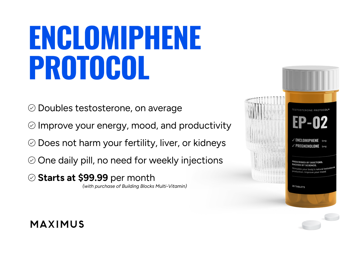
is "Enclomiphene Citrate” going to replace hCG in the USA?"

The discussion has touched on the effect of Clomid (Clomiphene, 60% enclomiphene & 40% zuclomiphene) and it would not seem to be a good way to go for a high SHBG because whilst Total Testosterone will increase, so will SHBG levels, resulting (at best) in only a small change inFree Testosterone values.

Regular IM/SubQ Testosterone would still see the way to go if hCG issues are no a factor?

If hCG factors are a concern then the thread above may be of interest and I draw your attention to the VIDEO found in the fist post of the thread, which started the debate.
 
Thanks, jacb, I just watched the video and will ask my doc about enclomiphene citrate. But I am doubtful it would work as monotherapy for me. Clomid monotherapy worked only on paper, raising my total T quite a bit without reducing my symptoms. I didn't have the side effects others find difficult, but it didn't work. I still essentially had low T because my free T remained low. I am currently trying Natesto monotherapy but am not long enough into it yet to judge whether it's working. My doc's basic approach is for me to try the things that will least shut down my body's own production of T. So he started with Clomid; now Natesto; and if Natesto doesn't work for me, then we would try a compounded cream.
 
In the recent years biomolecular interactions between T, SHBG, and magnesium have been studied by high performance liquid chromatography (HPLC) [92]. Excoffon and colleagues [92] provided evidence of a magnesium-mediated variation in the T-SHBG affinity. The change in magnesium levels inside the biological serum concentration range (0.75–0.95 mM) could lead to an enhancement of the Bio-T. In fact, the affinity of T to SHBG seems to change slightly with the magnesium concentration. Magnesium binds SHBG in a nonspecific mode, leading to an uncompetitive inhibition with T in binding SHBG and to a subsequent enhancement of Bio-T availability. The binding is accompanied by a magnesium release (or uptake) with a corresponding heat effect around in magnitude 17 kJ/mol [92].

 
Thanks, jacb, I just watched the video and will ask my doc about enclomiphene citrate. But I am doubtful it would work as monotherapy for me. Clomid monotherapy worked only on paper, raising my total T quite a bit without reducing my symptoms. I didn't have the side effects others find difficult, but it didn't work. I still essentially had low T because my free T remained low. I am currently trying Natesto monotherapy but am not long enough into it yet to judge whether it's working. My doc's basic approach is for me to try the things that will least shut down my body's own production of T. So he started with Clomid; now Natesto; and if Natesto doesn't work for me, then we would try a compounded cream.
Thanks Dudley

If I understand correctly … I believe that Low T Nation thinks that there is a fundamental difference between the effects of Clomid compared with the effects of taking only Enclomiphene. In reality the difference will be due to the missing effect of zuclomiphene found in Clomid.

You talk about mono therapy and natural T production. Some people used to try hCG mono therapy. But HCG is now hard to find and even when fully stimulated your body may not have produced enough T’?

Covid would have proved your potential to produce T’ ….. but it was not a good long term option because of the possible side effects and the fact that free testosterone was not increased because SHBG values were also elevated by the zuclomiphene.

Low T Nation are currently offering Enclomiphene to their clients saying that like Clomid it will elevate you natural T’ production to its maximum (which may or may not be sufficient) without significantly raising SHBG and because of that, you free T’ would also increase.

The jury is still out on what happens if you don’t produce enough T’ naturally while on Enclomiphene and wish to supplement with injected T’?
 
Thanks for this insight, Vince. What would you recommend I try?
 
Buy Lab Tests Online
Defy Medical TRT clinic

Sponsors

bodybuilder test discounted labs
cheap enclomiphene
TRT in UK Balance my hormones
Discounted Labs
Testosterone Doctor Near Me
Testosterone books nelson vergel
Register on ExcelMale.com
Trimix HCG Offer Excelmale
BUY HCG CIALIS

Online statistics

Members online
3
Guests online
5
Total visitors
8

Latest posts

Top