Nelson Vergel
Founder, ExcelMale.com

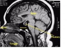
Pituitary imaging findings in male patients with hypogonadotrophic hypogonadism.
Authors
Hirsch D, et al. Pituitary. 2014 Sep 23. [Epub ahead of print]
Abstract
CONTEXT: Data on pituitary imaging in adult male patients presenting with hypogonadotrophic hypogonadism (HH) and no known pituitary disease are scarce.
OBJECTIVE: To assess the usefulness of pituitary imaging in the evaluation of men presenting with HH after excluding known pituitary disorders and hyperprolactinemia.
DESIGN: A historical prospective cohort of males with HH.
PATIENTS: Men who presented for endocrine evaluation from 2011 to 2014 with testosterone levels <10.4 nmol/L (300 ng/mL), normal LH and FSH levels and no known pituitary disease.
RESULTS: Seventy-five men were included in the analysis. Their mean age and BMI were 53.4 ± 14.8 years and 30.7 ± 5.2 kg/m(2), respectively. Mean total testosterone, LH, and FSH were 6.2 ± 1.7 nmol/L, 3.4 ± 2 and 4.7 ± 3.1 mIU/L, respectively. Prolactin level within the normal range was obtained in all men (mean 161 ± 61, range 41-347 mIU/L). Sixty-two men had pituitary MRI and 13 performed CT. In 61 (81.3 %) men pituitary imaging was normal. Microadenoma was found in 8 (10.7 %), empty sella and thickened pituitary stalk in one patient (1.3 %) each. In other four patients (5.3 %) a small or mildly asymmetric pituitary gland was noted. No correlation was found between testosterone level and the presence of pituitary anomalies.
CONCLUSIONS: This study suggests that the use of routine hypothalamic-pituitary imaging in the evaluation of IHH, in the absence of clinical characteristics of other hormonal loss or sellar compression symptoms, will not increase the diagnostic yield of sellar structural abnormalities over that reported in the general population.


