maxadvance
Active Member
So I ran across the term "Ventrogluteal IM injection site" and found an article describing it as what is currently the most medically recommended injection site. I tried it a few times now and have to agree. No blood gushing pinholes, no veins, no nerves, minimal pain.
IN: The Ventrogluteal IM injection site.
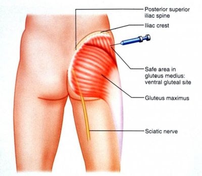
The ventorgluteal (VG) site has less subcutaneous fat and a thicker muscle mass than the dorsogluteal site with an almost certain probability of penetrating muscle with a standard needle.
The VG site is also sparse of any major innervating nerves or blood vessels whilst remaining well perfused from smaller branches.
Locating the VG site.
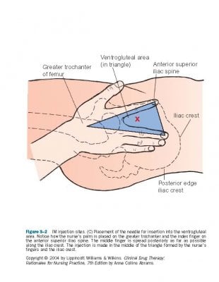
The ventrogluteal site is located halfway between the hip and the head of the femur. One method to locate the correct site is:
First, place the heel of your hand (use your L hand if injecting into the patients R VG and vice-versa) over the patients greater trochanter, and feel for the anterior superior iliac spine with your index finger.
The middle finger then slides across to make a peace-sign pointing up to the iliac crest.
The injection site is in the middle of this peace-sign.
Wipe site with alco-wipe in a circular motion and allow to dry.
Use your peace sign to spread skin taut.
Insert needle at 90 degree angle. Take care as you are inserting needle in proximity to your fingers.
There is no evidence for the need to aspirate the plunger when using the VG site.
Inject medication slowly (around 10 seconds per ml), remove needle quickly, and gently apply pressure to site for 10 seconds.
OUT: The Dorsogluteal IM injection site.
This site been used by nurses for years as the target of choice for IM injections.
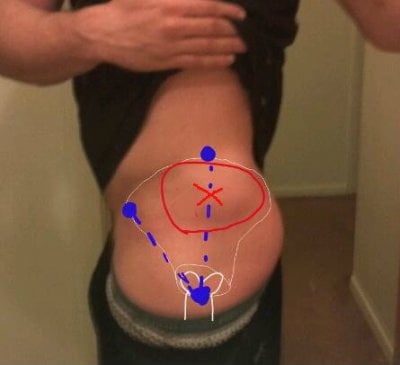
It is found in the area of the superior lateral aspect of the gluteal muscles, commonly known as the upper outer quadrant.
It is located by dividing the buttock into four equal quadrants. This is usually done by drawing an imaginary cross (bisecting it vertically and horizontally).
Problems that have been identified with using this site include:
Presence of major nerves and blood vessels in this area, including the sciatic nerve and superior gluteal artery.
It has been taught that you will probably avoid this by further dividing the upper outer quadrant into another quadrant and giving the injection into the upper outer of the upper outer.
Despite this, there have been reports of injuries to the sciatic nerve leading to problems ranging from foot drop to paralysis of the lower limb.
Thickness of fat in this area. A number of studies have found that the depth of muscle in the dorsogluteal region is often greater then the length of a standard needle used for IM injections, resulting in a failure to achieve intramuscular deposition of the medication.
In fact, one study found the success rate of IM injections to be 32% (which fell to 8% in female patients)!
With the increasing incidence of obesity amongst our patients we are probably going to be delivering subcutaneous injections if we choose this location.
Pain receptors are located in the subcutaneous layer, not in muscle tissues and so medication delivered into this area may be more painful.
Dorsogluteal site has a decreased absorption rate increasing the possibility of a depot effect with drug build up and potential for overdose.
Still IN: the Z-track.

When delivering medications via any IM route the technique of Z-tracking should be used.
This both reduces pain, and prevents dispersion of medication into subcutaneous tissue.
Apply gentle traction on the skin to pull it away from the injection site (about 2-3 cm). Use your non-dominant hand.
Inject (slowly) with needle at 90 degrees to skin surface.
Withdraw needle quickly.
Release skin.
Summary:
Whenever possible the VG site should be the preferred location for intramuscular injections.
The Z-Track method should be used for IM medication delivery.
The patient should be positioned so the target muscle is as relaxed as possible.
IN: The Ventrogluteal IM injection site.
The ventorgluteal (VG) site has less subcutaneous fat and a thicker muscle mass than the dorsogluteal site with an almost certain probability of penetrating muscle with a standard needle.
The VG site is also sparse of any major innervating nerves or blood vessels whilst remaining well perfused from smaller branches.
Locating the VG site.
The ventrogluteal site is located halfway between the hip and the head of the femur. One method to locate the correct site is:
First, place the heel of your hand (use your L hand if injecting into the patients R VG and vice-versa) over the patients greater trochanter, and feel for the anterior superior iliac spine with your index finger.
The middle finger then slides across to make a peace-sign pointing up to the iliac crest.
The injection site is in the middle of this peace-sign.
Wipe site with alco-wipe in a circular motion and allow to dry.
Use your peace sign to spread skin taut.
Insert needle at 90 degree angle. Take care as you are inserting needle in proximity to your fingers.
There is no evidence for the need to aspirate the plunger when using the VG site.
Inject medication slowly (around 10 seconds per ml), remove needle quickly, and gently apply pressure to site for 10 seconds.
OUT: The Dorsogluteal IM injection site.
This site been used by nurses for years as the target of choice for IM injections.
It is found in the area of the superior lateral aspect of the gluteal muscles, commonly known as the upper outer quadrant.
It is located by dividing the buttock into four equal quadrants. This is usually done by drawing an imaginary cross (bisecting it vertically and horizontally).
Problems that have been identified with using this site include:
Presence of major nerves and blood vessels in this area, including the sciatic nerve and superior gluteal artery.
It has been taught that you will probably avoid this by further dividing the upper outer quadrant into another quadrant and giving the injection into the upper outer of the upper outer.
Despite this, there have been reports of injuries to the sciatic nerve leading to problems ranging from foot drop to paralysis of the lower limb.
Thickness of fat in this area. A number of studies have found that the depth of muscle in the dorsogluteal region is often greater then the length of a standard needle used for IM injections, resulting in a failure to achieve intramuscular deposition of the medication.
In fact, one study found the success rate of IM injections to be 32% (which fell to 8% in female patients)!
With the increasing incidence of obesity amongst our patients we are probably going to be delivering subcutaneous injections if we choose this location.
Pain receptors are located in the subcutaneous layer, not in muscle tissues and so medication delivered into this area may be more painful.
Dorsogluteal site has a decreased absorption rate increasing the possibility of a depot effect with drug build up and potential for overdose.
Still IN: the Z-track.
When delivering medications via any IM route the technique of Z-tracking should be used.
This both reduces pain, and prevents dispersion of medication into subcutaneous tissue.
Apply gentle traction on the skin to pull it away from the injection site (about 2-3 cm). Use your non-dominant hand.
Inject (slowly) with needle at 90 degrees to skin surface.
Withdraw needle quickly.
Release skin.
Summary:
Whenever possible the VG site should be the preferred location for intramuscular injections.
The Z-Track method should be used for IM medication delivery.
The patient should be positioned so the target muscle is as relaxed as possible.
Last edited by a moderator:


