madman
Super Moderator
Allosterically coupled multi-site binding of testosterone to human serum albumin
Abhilash Jayaraj, Heidi A. Schwanz, Daniel J. Spencer, Shalender Bhasin, James A. Hamilton, B. Jayaram, Anna L. Goldman, Meenakshi Krishna, Maya Krishnan, Aashay Shah, Zhendong Jin, Eileen Krenzel, Sashi N. Nair, Sid Ramesh, Wen Guo, Gerhard Wagner, Haribabu Arthanari, Liming Peng, Brian Lawney, Ravi Jasuja
ABSTRACT
Human serum albumin (HSA) acts as a carrier for testosterone, other sex hormones, fatty acids, and drugs. However, the dynamics of testosterone's binding to HSA and the structure of its binding sites remain incompletely understood. Here, we characterized the dynamics of testosterone's binding to HSA and the stoichiometry and structural location of the binding sites using two-dimensional nuclear magnetic resonance (2D NMR), fluorescence spectroscopy, bis-ANS partitioning, and equilibrium dialysis, complemented by molecular modeling.
2D NMR studies showed that testosterone competitively displaced 18-[ 13C]- oleic acid from at least three known fatty acid-binding sites on HSA that also bind many drugs. Binding isotherms of testosterone's binding to HSA generated using fluorescence spectroscopy and equilibrium dialysis were nonlinear and the apparent Kd varied with different concentrations of testosterone and HSA. The binding isotherms neither conformed to a linear binding model with 1:1 stoichiometry nor to two independent binding sites; the binding isotherms were most consistent with two or more allosterically coupled binding sites. Molecular dynamics studies revealed that testosterone's binding to fatty acid-binding site 3 on HSA was associated with conformational changes at site 6, indicating that residues in these two distinct binding sites are allosterically coupled.
Conclusion
There are multiple, allosterically coupled binding sites for testosterone on HSA. Testosterone shares these binding sites on HSA with free fatty acids, which could displace testosterone from HSA under various physiological states or disease conditions, affecting its bioavailability.
INTRODUCTION
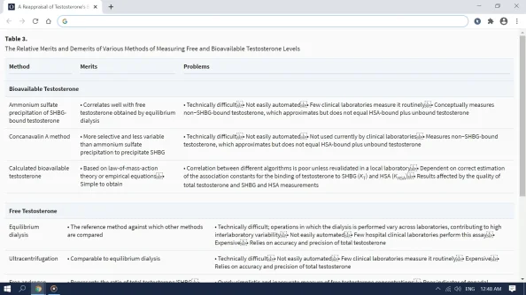
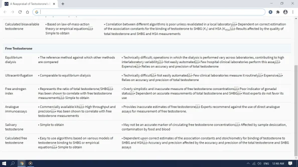
Human serum albumin (HSA) is the most abundant protein in human plasma, with concentrations ranging from 30-50 g·L-1 (approximately 450-750 µM). HSA acts as a carrier for endogenous lipophilic compounds such as steroid hormones, fatty acids, some nutrients, and many drugs (1, 2). The steroid hormones, testosterone (T), dihydrotestosterone (DHT), and estradiol (E2), bind reversibly with high affinity to sex hormone-binding globulin (SHBG) and with lower affinity to HSA. Because of relatively high HSA concentrations, a substantial (33 to 54%) fraction of testosterone in the plasma is carried by HSA. Total testosterone levels represent the sum of the concentrations of protein-bound and unbound (free) testosterone in circulation. Free testosterone refers to the fraction of circulating testosterone that is not bound to any circulating protein while the bioavailable fraction refers to the circulating testosterone that is not bound to SHBG, reflecting the view that HSA-bound testosterone can dissociate from HSA at the capillary level, especially in tissues with long transit time (due to low affinity), and may be available for biological action. Laurent et al have shown that binding of testosterone to SHBG affects its bioavailability and that the biological activity of circulating testosterone correlates with the free testosterone levels (3-5). The binding to SHBG and HSA regulates the distribution of circulating testosterone into its bound and free fractions. HSA, even with its relatively lower affinity than SHBG, binds a higher fraction of circulating testosterone in men and women than SHBG due to its high binding capacity and high concentration. However, the dynamics of testosterone's binding to HSA remain incompletely understood and were the subject of this investigation.
Structurally, HSA is comprised of three homologous domains (6); domains II and III both contain a binding pocket formed mostly of hydrophobic and positively charged residues in which a variety of compounds bind (7–15). Although it has been shown that HSA possesses multiple binding sites for several biomolecules, including fatty acids and some drugs, it is generally believed that testosterone binds to HSA at a single site on domain IIA (10–12) with a low-to-moderate affinity (Ka ≈ 2.0 − 4.1 × 10⁴ M -1 at 37°C) (13–18) and 1:1 stoichiometry. Because HSA transports a number of hormones, nutrients, and exogenous drugs, these endogenous ligands and drugs could potentially compete with testosterone for the same binding pocket(s) on HSA, thereby affecting its binding and bioavailability.
Since the publication of the crystal structure of HSA by He and Carter in 1992 (19), structural aspects of the binding sites of HSA in its physiologically relevant solution state have been characterized using nuclear magnetic resonance (NMR). Our group pioneered the novel strategy of using 13C-enrichment of a specific carbon in a fatty acid as a non-perturbing probe to enable visualization of small ligands complexed with a large protein, for which a solution structure has not been obtained (20). Since then, the use of two-dimensional (2D) NMR of 13C-enriched fatty acids has facilitated the identification of common binding sites for many biomolecules and drugs on HSA. The application of 2D NMR to characterize 18-[ 13C]-oleic acid (OA) binding to HSA led to the identification of nine individual OA binding sites within the three domains of HSA; seven of these nine binding sites have since been mapped onto the crystal structure (19). Some of these fatty acid-binding sites also participate in binding other biomolecules and drugs (6), each of which can competitively alter the binding of the other ligands (22, 23). OA bound to three low-affinity sites can be displaced by drugs from Sudlow’s drug sites I and II, as well as from the fatty acid-binding site 6 (23). Along similar lines, free fatty acids have been shown to modulate the binding of steroid hormones, including testosterone, to HSA, (24-26) leading us to consider the hypothesis that testosterone binds to one or more of the fatty acid-binding sites on HSA. Accordingly, we utilized the well-characterized HSA: OA 2D NMR system, described above, to investigate the competitive displacement of 18- [ 13C]-oleic acid by testosterone and determine the stoichiometry and the structural/spatial location of the testosterone binding pocket(s) on HSA.
Because chemical shift perturbations detected by NMR can be caused by either a direct binding event in the immediate binding pocket or by allosteric coupling distant from the binding site, we performed molecular dynamics studies to characterize the binding affinities of each site for testosterone and OA and explain the sequence of displacement and binding observed experimentally. Structural perturbations in HSA binding regions distant from the immediate binding site caused by the binding of testosterone to a specific binding site were evaluated by the following root mean square fluctuations (RMSF), residue cross-correlations, and principal component analysis (PCA). Additionally, the findings of the 2D NMR experiments were confirmed by studying the binding dynamics of testosterone using two independent methods: steady-state fluorescence spectroscopy and equilibrium dialysis. Collectively, the data reported in this manuscript on the chemical shift perturbation observed in the 2D NMR studies; the results of the fluorescence spectroscopy and equilibrium dialysis experiments; and the molecular modeling provide important insights into the locations of the multiple binding sites of testosterone on HSA, the dynamics of binding pocket residue coupling in the HSA: testosterone complex, and the molecular understanding of the binding stoichiometry.
DISCUSSION
Here, we employed multiple biophysical techniques – 2D NMR, fluorescence spectroscopy, bis-ANS partitioning, and equilibrium dialysis experiments – along with complementary molecular modeling studies to characterize the stoichiometry and the structure of the testosterone binding pockets on HSA. Our 2D NMR data offer direct experimental evidence that testosterone binds to at least three FA binding sites on HSA and the steady-state fluorescence quenching of tryptophan residues and repartitioning of bis-ANS probe show that the testosterone binding to HSA is a multiphasic process. The detailed molecular modeling studies suggest that these binding events are associated with significant conformational rearrangement in the binding pockets – distant from the site of testosterone's binding – indicating that they are allosterically coupled. These data do not support the prevailing model of 1:1 stoichiometry of testosterone's binding to a single binding site on HSA. These findings are novel and significant in several aspects. First, they show that there are multiple binding sites for testosterone on HSA (the prevalent view is that testosterone binds to HSA at a single binding site with a single Kd). Second, we show for the first time that these binding sites are allosterically-coupled. Finally, our data show for the first time that testosterone shares these binding sites on HSA with free fatty acids and some commonly used drugs and provide a mechanistic explanation for how commonly used drugs and free fatty acids – especially in the postprandial state and in disease conditions characterized by elevated free FA concentrations – can displace testosterone from its binding sites on HSA and potentially affect its bioavailability.
The 2D NMR data suggest the presence of at least three testosterone binding sites on HSA and the fluorescence spectroscopy data showed that these binding sites have distinct binding affinities. Equilibrium dialysis experiments revealed that the apparent Kd changes depending on the ratio of the concentrations of testosterone and HSA, providing further evidence of two or more binding sites and the possibility of allosteric coupling between the sites. The molecular modeling results confirmed the presence of allosterically coupled testosterone binding sites and provided novel insights into the energetics of the binding and the relative affinities of the binding sites for testosterone. The cross-correlation matrices provided confirmation of the interaction between the sites participating in testosterone binding.
The two Kd values resulting from the fit of the steady-state data (Figure 3B) are 17.8 nM and 12.3 µM, which differ by three orders of magnitude. The range of circulating testosterone concentration in men is 10-35 nM, that of HSA 450-750 µM, and that of SHBG 13.5-87.3 nM. With such high serum concentration of HSA, comparatively lower concentrations of testosterone and SHBG, and a high-affinity binding pocket on HSA in the low nanomolar range, virtually all of the testosterone in the blood would expect to be bound to HSA, with a negligible amount bound to SHBG or unbound to any protein. However, clinical data (13, 18, 48) show that 33- 45% of testosterone in the blood is bound to SHBG, while 50-67% is bound to HSA and 1-4% is unbound. The only explanation for this, while staying consistent with the derived dissociation constant values from our data, is that the low-affinity site with a Kd of 12.3 µM is the only available site on HSA in the unbound state. The binding of testosterone to the low-affinity binding site causes a long-range conformational change that renders the second, high-affinity binding site available for binding testosterone. We have previously observed similar allosteric coupling between the monomers of dimeric SHBG upon testosterone binding (49). The overly simplified linear models with 1:1 stoichiometry have overlooked the complexities in the dynamics of the binding of testosterone to HSA and SHBG.
CONCLUSION
Using 2D NMR, equilibrium dialysis, fluorescence spectroscopy, and molecular modeling, we provide evidence of at least three allosterically coupled binding sites for testosterone on HSA. We show that testosterone binds to the same binding sites that are known to bind with fatty acids (FAs) and other endogenous ligands, and many commonly used drugs. These data also provide a mechanistic explanation for how commonly used drugs and free FAs – especially in the postprandial state and in disease conditions characterized by elevated free FA concentrations – could displace testosterone from its binding sites on HSA and potentially affect its bioavailability.


